Dr. Tricia and Michael Pirone pride themselves on having the most advanced technology in Eyecare. They strive to diagnose,prevent, treat and educate their patients concerning the health and wellness of their vision.
Glaucoma is a neurodegenerative disease that can lead to blindness due to loss of retinal ganglion cells. With the RTVue OCT, we are able to detect if a patient has glaucoma. The RTVue examines the macular ganglion cell layer, and gives a 3D scan of the optic nerve. It also measures the corneal thickness.
The OCT allows us to evaluate a patient’s retinal condition by taking many cross sections of the retina. The retinal scan allows us to diagnose and monitor conditions such as macular degeneration, diabetic or hypertensive retinopathy, retinal holes or detachments, choroidal nevi, and central serous retinopathy.
The doctors at Eyes On Rosemont have been using the TRS 5100 since they opened in 2009. This is an automated refracting system that replaces the standard refractor. This system helps generate an extremely accurate prescription for the patient, is very patient friendly, and quickly integrates into the patients electronic medical record.
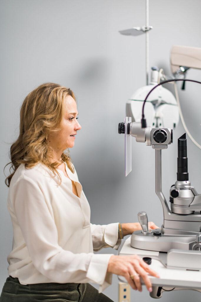
Nidek automatic refractor and keratometer allows a rapid accurate testing for proper prescription. The doctor follows this verification by refraction to customize your Rx to the proper focus.
This is a quick clinical diagnostic test that we do on all patients. This can detect visual field loss that can indicate glaucoma or other ocular or neurological disorders. It can detect glaucoma earlier than eye pressure testing alone.
This is the gold standard in perimeter. Validated by more than 30 years of research, design and clinical experience, the Humphrey Field Analyzer is the accepted standard of care in glaucoma diagnosis and management.
The Icare tonometer is based on a new measuring principle, in which a very light probe is used to make momentary contact with the cornea. The measurement is barely noticed by the patient.
The Icare rebound tonometer is extremely efficient and accurate at measuring pressures in the eye. It is fast, requires no drops, and requires no puff of air. Most of our patients love this device and is now our go-to method of iop measurement.
This checks eye pressure without any puff. It is a quick, easy, and accurate way of measuring the pressure in the eye.
The Optus Daytona is a scanning retinal imaging instrument that is able to take ultra-wide images of a patient’s retina. This device can take around a 200-degree image of the retina in one image and can capture the picture in less than one second.
This measures patients’ Macular Pigment Optical Density. There are three carotenoids that comprise the macular pigment: Zeaxanthin, Lutein, and meso-Zeaxanthin. It is believed that healthy amounts of macular pigment help block harmful blue light, acting as “internal sunglasses”, help prevent age related macular degeneration, and increase visual performance.
The OCULUS Keratograph 5M is an advanced corneal topographer with a built in real keratometer and a color camera optimized for external imaging. This provides extremely precise details about the surface characteristics of the entire cornea. Circular patterns of light are beamed into your eye for analysis. A computer creates and prints out the resulting surface “map” of your eye, with different contours represented by varying colors. The OCULUS also has a unique feature in that it examines the meibomian glands, the tear film break-up time, the tear meniscus height and evaluates the lipid layer of the tears.
We are thrilled to welcome Dr. Allie Decker and Dr. Sydney Butalla to our team! Now offering expert eyecare for kids of all ages-from the ages of 6 months to teens. Book your appointment today! 207-775-1819 – 207-210-6700